ULTRASOUND IMAGING
Discover the power of clarity with our advanced point of care ultrasound imaging, enhancing accuracy and insight in podiatric practice and injury rehabilitation.

Services
WHAT IS POINT OF CARE ULTRASOUND IMAGING?
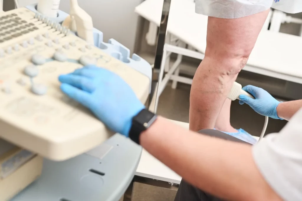
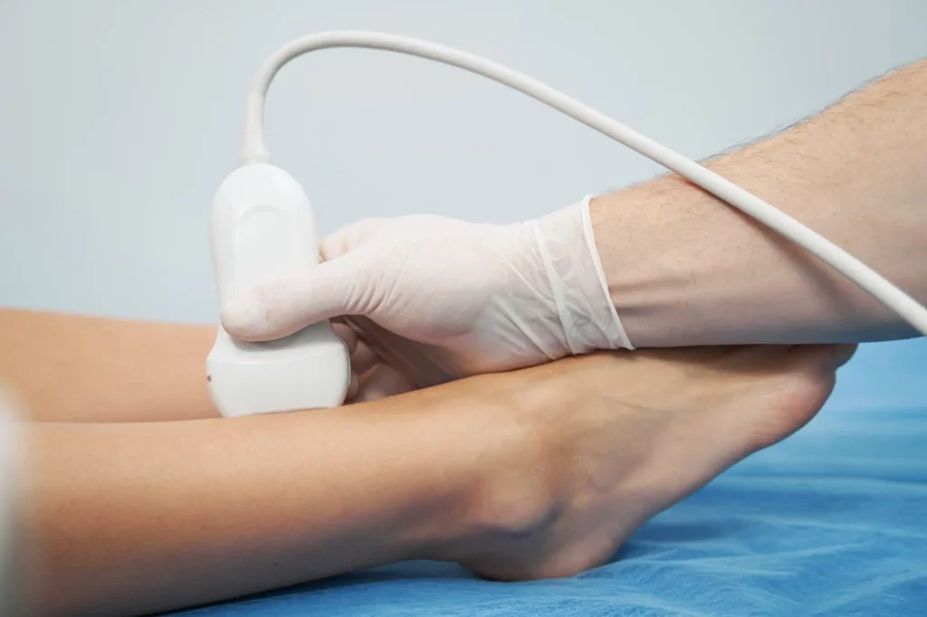
Musculoskeletal (MSK) Point of Care Ultrasound (POCUS) imaging is a non-invasive, real-time diagnostic tool used to visualise internal structures within the body, such as: muscles, tendons, ligaments, and bones. Specifically, within podiatry, POCUS allows for detailed imaging of the calf, foot and ankle, aiding in the assessment and diagnosis of various conditions.
Key Features of POCUS in Podiatry:
Real-time Visualisation:
POCUS provides instant visual feedback, allowing podiatrists to observe dynamic changes to structures in the foot and ankle during movement.
Safe:
Unlike traditional imaging methods, POCUS is non-invasive and uses soundwaves rather than radiation to produce images. Making it a comfortable and patient-friendly diagnostic option.
Precision and Detail:
The high-resolution images produced by POCUS enable the identification of subtle anatomical variations and pathologies to muscles, tendons, ligaments and bones within the foot and ankle.
Informed Decision Making:
By offering immediate visual information, POCUS empowers podiatrists to make timely and accurate clinical decisions, leading to more focused treatment plans.
WHAT IS MSK POINT OF CARE ULTRASOUND IMAGING USED FOR?
At MyFootMedic, Point of Care Ultrasound (POCUS) is used as a powerful tool for evaluating and helping treat a range of foot and ankle conditions, in conjunction with a physical assessment, such as:
Soft Tissue Injuries: POCUS helps us see damage to the soft parts of your body, like muscles and tendons, so we can choose the best way to help you heal.
Ligament and Tendon Disorders: By using POCUS, we can spot problems with your ligaments and tendons early, making it easier to plan your treatment.
Joint Problems: POCUS lets us take a close look at the joints in your feet and ankles, helping us understand and address any issues you might be having.
Nerve Entrapments: POCUS assists us in identifying places where your nerves might be pinched or trapped, guiding us in finding the right solutions for you.
By using POCUS for these conditions, we can provide you with more precise and effective care, tailored to your specific needs.
HOW WILL POINT OF CARE ULTRASOUND IMAGING HELP ME?
When you visit MyFootMedic, our use of Point of Care Ultrasound (POCUS) will benefit you in several important ways:
Clear Understanding:
POCUS allows you to see what's happening inside your feet and ankles, helping you and our team gain a clearer understanding of your condition.
Quick Answers:
By providing immediate imaging, POCUS helps us swiftly identify the issue, meaning you spend less time waiting for answers and more time focusing on your treatment.
Tailored Treatment:
With the detailed insights from POCUS, we can create a personalised treatment plan that is specifically designed to address your unique needs.
Minimised Discomfort:
POCUS is a non-invasive and painless imaging technique, ensuring your comfort during the diagnostic process.
Informed Decisions:
The real-time images produced by POCUS enable us to make well-informed decisions about your care, leading to more effective and timely treatment.
By incorporating POCUS into your care, we aim to provide you with a comprehensive and transparent approach that prioritises your well-being and recovery.
HOW DOES POINT OF CARE ULTRASOUND IMAGING WORK?
At MyFootMedic, Point of Care Ultrasound (POCUS) works by using high-frequency sound waves to create detailed images of the inside of your calf, feet and ankles. Here’s how it works:
1
Sound Wave Transmission:
2
Reflection of Sound Waves:
3
Image Formation:
4
Dynamic Visualization:
By harnessing the power of POCUS, we can gain a deeper understanding of your foot health, leading to more accurate diagnoses and tailored treatment plans.
Our clinic only utilises POCUS for injury and MSK structure assessments, we do not perform POCUS assessments for cancer detection or vascular assessments. We specialise in MSK assessments of the calf, ankle and feet, we do not perform POCUS scans for other areas of the body.
LET’S ANSWER YOUR POINT OF CARE ULTRASOUND IMAGING QUESTIONS!
Point-of-care ultrasound (POCUS) and normal ultrasound differ primarily in the user and location. A normal diagnostic ultrasound is typically performed by a sonographer or radiologist in a dedicated imaging department, focusing on comprehensive imaging assessments of specific body areas without performing physical assessments. In contrast, POCUS is conducted by practitioners (e.g. Podiatrists, Physiotherapists, GP’s) in conjunction with a physical examination, to obtain real-time information for immediate clinical decision-making.
Point of Care Ultrasound (POCUS) is generally considered to be highly accurate when performed by trained healthcare professionals. POCUS is known for its reliability in quickly providing valuable diagnostic information, aiding in timely decision-making. Overall, when used by skilled practitioners, POCUS is a valuable tool for rapid and accurate diagnostic assessment in various clinical settings.
A foot point of care ultrasound is a non-invasive imaging test that can provide detailed visuals of the bones, joints and soft tissues in the foot, such as tendons, ligaments, muscles, and nerves. It can help diagnose conditions like tendonitis, plantar fasciitis, or ligament tears, as well as identify any abnormalities or fluid accumulation or degenerative changes to joints. Overall, a foot ultrasound is valuable for evaluating a wide range of foot and ankle problems, aiding in accurate diagnosis and treatment planning.
The duration of a scan can vary depending on the specific circumstances and the reason for the procedure. Generally, a foot ultrasound takes approximately 10-15 minutes to complete. However, this timeframe may be longer if there are complications or if a more detailed examination is required.
Yes, point of care ultrasound imaging can be used to detect Morton’s neuroma. Ultrasound imaging is a non-invasive and effective method for visualising this condition. It can accurately locate the neuroma, assess its size, and help differentiate it from other conditions causing foot pain. Additionally, ultrasound is useful for guiding therapeutic injections and can aid in treatment planning for Morton’s neuroma.
Yes, plantar fasciitis can be detected using point of care ultrasound imaging. Ultrasound is a commonly used and effective tool for diagnosing plantar fasciitis as it can reveal thickening, swelling, and other abnormalities in the plantar fascia. It also allows for real-time visualisation of the affected area, making it a valuable diagnostic aid for healthcare professionals. If you are experiencing symptoms of plantar fasciitis, consulting a podiatrist for a proper diagnosis and treatment plan is recommended.
POINT OF CARE ULTRASOUND IMAGING IN BEDFORD WITH MY FOOT MEDIC.
Discover the benefits of Point of Care Ultrasound (POCUS) imaging for foot and ankle concerns at our Bedford facility. Our experienced podiatrists and advanced technology ensure effective diagnosis and personalised treatment plans.
Contact us today to schedule an appointment and experience the advantages of ultrasound imaging in podiatric care.


